
obstetrics
Ultrasound: How It Works, What It Can See, and Whether It Is Safe
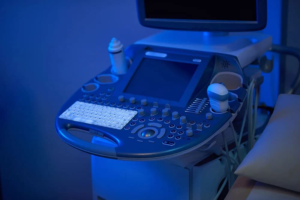
Ultrasound is the workhorse of obstetrics and gynecology. It uses high-frequency sound waves rather than ionising radiation, which is why it can be repeated through a pregnancy without the dose accounting an X-ray or CT scan requires.
This guide covers how it works, the types you may be offered, what a scan can genuinely detect and what it cannot, and a direct answer on safety — because there is a specific piece of misinformation on this topic that frightens people away from care they need.
Is ultrasound safe in pregnancy? The short answer
Yes, when it is medically indicated and performed by a trained operator. Diagnostic ultrasound has been in routine obstetric use for over fifty years. ACOG, the American Institute of Ultrasound in Medicine (AIUM), and the FDA all consider it safe for that purpose. No study has demonstrated harm to a fetus from diagnostic-intensity ultrasound.
There is no established link between prenatal ultrasound and autism, speech delay, or learning problems. You will find this claim repeated across the internet — an earlier version of this very article repeated it, which is why we are correcting it explicitly rather than quietly deleting it. It is not supported by the evidence. Large studies have specifically looked for an association with autism spectrum disorder and have not found one. If anything, some research has found the opposite of what the claim predicts.
Two real caveats, which are about use rather than about the technology:
- Keepsake and "boutique" scans are not recommended. The FDA and AIUM both advise against non-medical fetal imaging — commercial 3D/4D souvenir sessions and at-home fetal dopplers. The objection is not that a scan is dangerous; it is that ultrasound deposits energy in tissue, so exposure should have a medical reason behind it and be kept to the shortest time and lowest power that answers the clinical question (the ALARA principle). An untrained operator satisfies none of that, and may also produce false reassurance.
- Therapeutic ultrasound is a different thing entirely. The tissue-heating and cavitation effects sometimes cited as "ultrasound risks" belong to high-intensity focused ultrasound used to treat tissue, not to diagnostic imaging.
How ultrasound works
A hand-held transducer converts electrical energy into sound waves and sends them into the body. The waves reflect off boundaries between tissues of different density and return to the transducer, which converts them back into electrical signals. A computer times those echoes and builds an image from them. Gel between the transducer and the skin removes the air gap that would otherwise stop the sound entering the body at all.
Diagnostic ultrasound uses frequencies between roughly 2 and 18 MHz — far above human hearing. Frequency is a trade-off:
- Higher frequency gives finer resolution but penetrates less. Used for superficial structures: thyroid, breast, and transvaginal scanning.
- Lower frequency penetrates deeper at the cost of detail. Used for abdominal imaging and for patients where the target is further from the probe.
Types of ultrasound you may be offered
- Transabdominal — the probe moves across the abdomen. The standard for most of pregnancy.
- Transvaginal — a slim probe is placed in the vagina, putting it much closer to the uterus and ovaries. Standard in early pregnancy and for gynecologic assessment, because the image quality is far better than a transabdominal view at that distance. It is not painful, though it can be uncomfortable, and you can ask for it to stop at any point.
- Doppler — measures the speed and direction of blood flow. Used to assess umbilical and fetal circulation, and to look for clots or vessel narrowing.
- 3D and 4D — reconstructs a surface image; 4D adds motion. Clinically useful for specific questions such as facial or skeletal anomalies. Most of its everyday fame is cosmetic.
- Fetal echocardiography — a detailed study of the fetal heart, offered when there is a risk factor or when a screening scan raises a question.
- Sonohysterography — sterile saline is placed in the uterine cavity to outline polyps, fibroids, or adhesions that a plain scan can miss.
Scans in pregnancy, and what each one is for
- First trimester (about 6–10 weeks) — confirms the pregnancy is in the uterus, confirms cardiac activity, counts the fetuses, and dates the pregnancy. Early dating by crown-rump length is the most accurate dating you will ever get; every later due-date decision rests on it.
- Nuchal translucency (11+0 to 13+6 weeks) — measures fluid at the back of the fetal neck as part of combined screening.
- Anatomy scan (18–22 weeks) — the detailed survey of fetal structures, placental position, and amniotic fluid. This is usually when fetal sex can be identified reliably, if you want to know.
- Third trimester, as indicated — growth, fluid volume, and fetal position where there is a reason to check.
What ultrasound can and cannot detect
This distinction causes more anxiety than any other part of the subject, so it is worth being precise.
Ultrasound can detect structural anomalies — spina bifida and other neural tube defects, many congenital heart defects, limb and abdominal wall abnormalities, kidney anomalies — plus placental position, growth restriction, and amniotic fluid problems.
Ultrasound cannot diagnose Down syndrome or any other chromosomal condition. It can only identify soft markers and measurements, such as nuchal translucency, that shift the calculated probability. A diagnosis requires NIPT (a screening blood test with high but not perfect accuracy) followed by CVS or amniocentesis, which examine fetal chromosomes directly and are the only diagnostic tests. A scan that "looks normal" does not exclude a chromosomal condition, and a soft marker does not establish one.
Detection is never complete. Even an excellent anatomy scan detects a proportion of structural anomalies, not all of them. Image quality is affected by fetal position, gestational age, abdominal wall thickness, and scar tissue — and it depends heavily on the operator's skill and the equipment.
Ultrasound outside pregnancy
In gynecology, ultrasound is the first-line imaging test for:
- Fibroids, endometrial polyps, and adenomyosis
- Ovarian cysts, and features that help distinguish benign from concerning ones
- Endometrial thickness, particularly in postmenopausal bleeding
- IUD position, when strings cannot be located
- Investigation of pelvic pain, abnormal bleeding, and infertility
It is also used well beyond this specialty — cardiology, abdominal imaging, musculoskeletal assessment, and to guide needles safely during biopsies and aspirations.
Limitations worth knowing
- Less effective than MRI or CT for some questions, particularly small soft-tissue lesions and anything inside the adult skull.
- Highly operator-dependent. Who performs and interprets the scan matters more than the machine's brand.
- Bowel gas and body habitus can obscure the view; a transvaginal approach often solves the pelvic version of this problem.
- False positives happen. A finding that turns out to be nothing can still cost you weeks of worry and further tests — a real harm, and a reason scans should answer a question rather than be done for their own sake.
Ultrasound at the Women's Health Center of Chicago
We perform obstetric and gynecologic ultrasound in our own offices, so you are scanned by the practice that is managing your care rather than sent elsewhere and left waiting for a report.
- Pregnancy and prenatal care
- Gynecological services
- Meet Dr. Adeeb Alshahrour
- Book an appointment online
Medical disclaimer
This article is general health information, not medical advice for any individual. What scans you need, and when, is a decision for you and your own clinician based on your pregnancy and your history. If a scan has raised a question about your pregnancy, talk it through with your provider rather than with a search engine.